Visual System Keynote Shapes: Detailed Eye Anatomy Visuals


Type: Keynote Shapes template
Category: Medicine - Pharma
Sources Available: .key
Product ID: KS00026
Template incl.: 14 editable slides
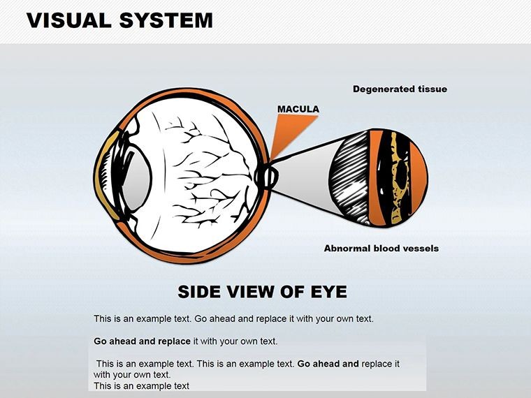
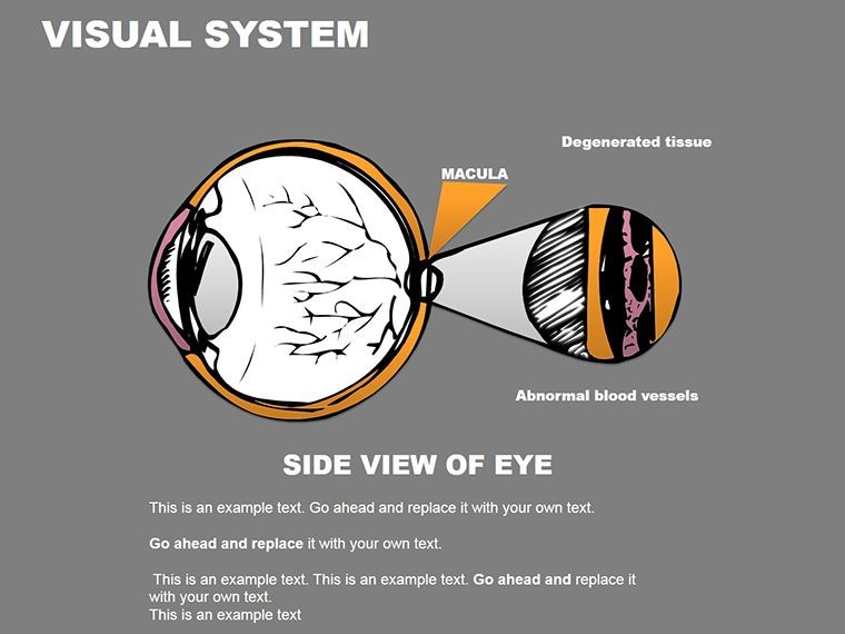
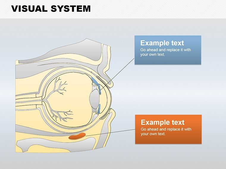
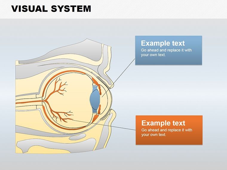
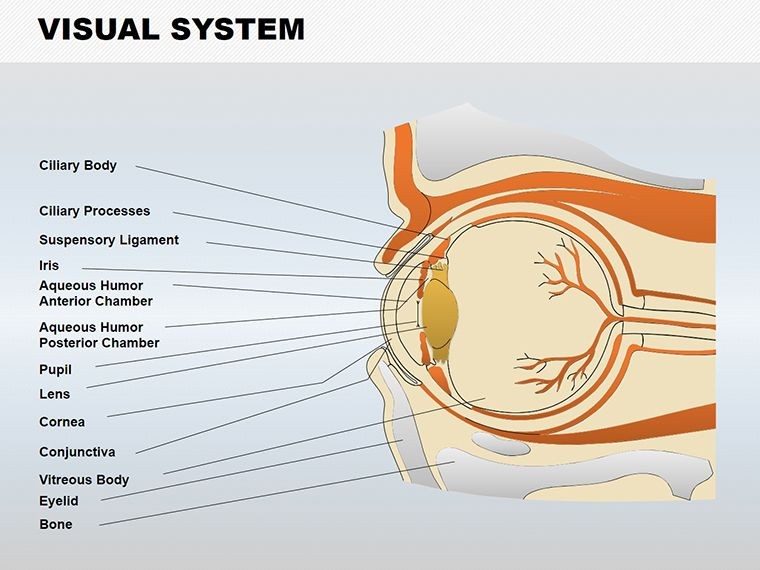
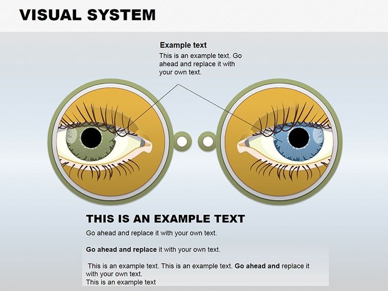
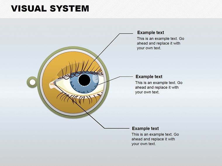
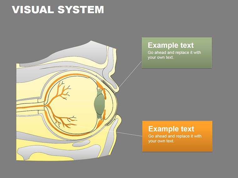
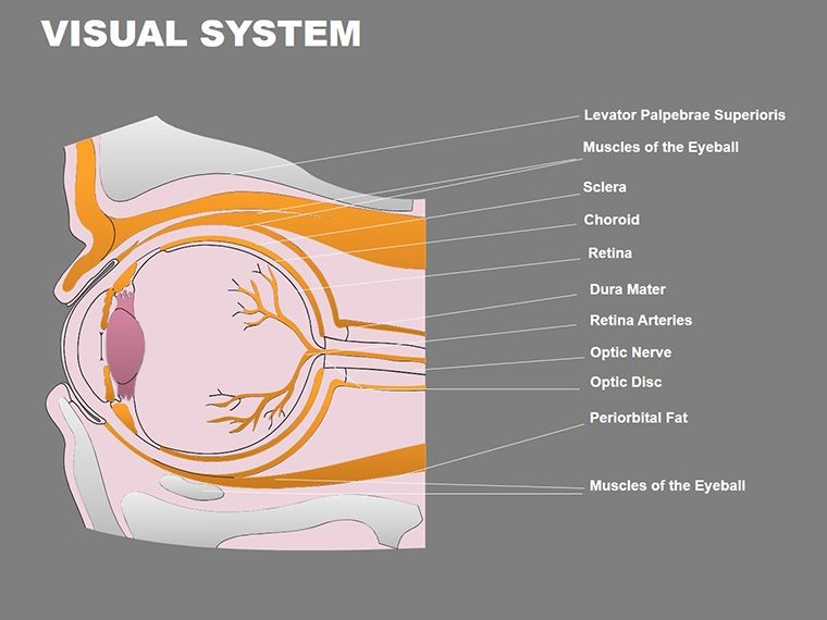
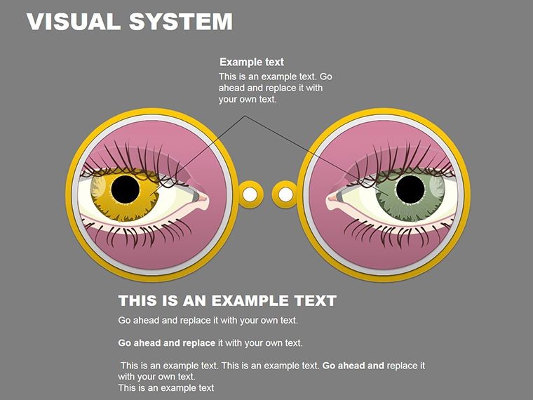
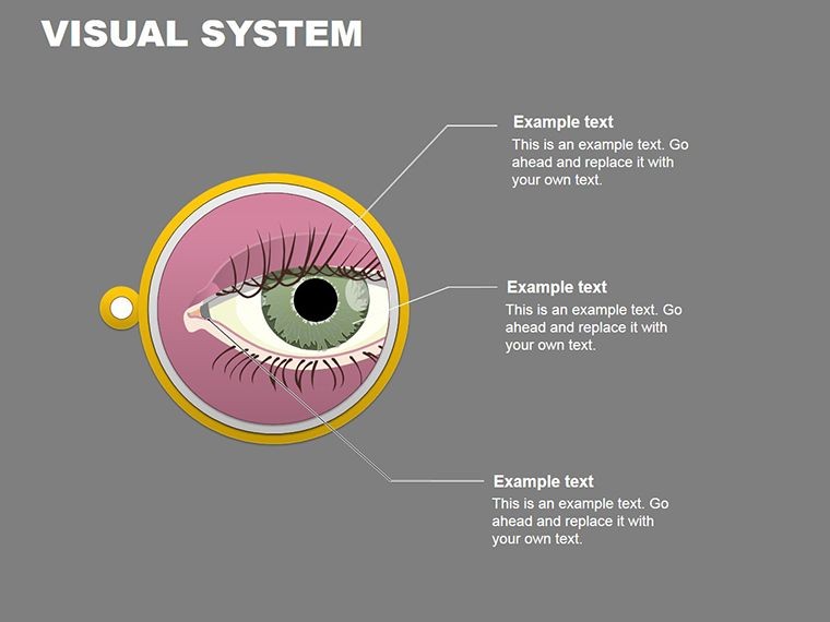
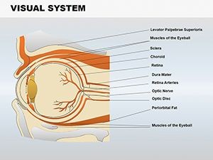
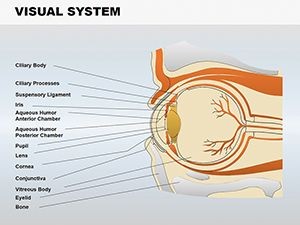
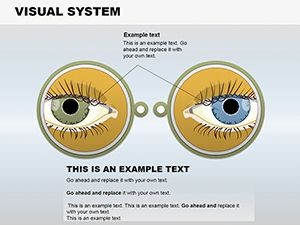
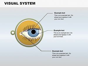
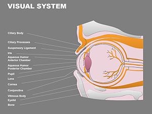
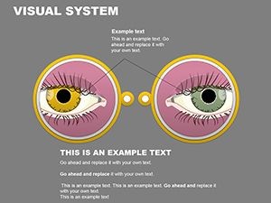
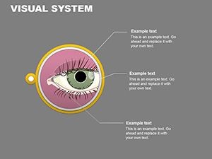
The human eye, a marvel of biology with its 2.5 cm diameter and intricate layers, deserves precise visualization in educational and medical contexts. Our Visual System Keynote shapes template provides 14 editable slides to detail components like the sclera, cornea, choroid, iris, and pupil. Tailored for healthcare professionals, educators, and researchers, it simplifies explaining light refraction (75% via cornea) or pupil dynamics in varying light. Compatible with Keynote (.key), it offers seamless edits for accurate representations. Informed by anatomical standards from sources like the American Academy of Ophthalmology, this tool enhances teaching and consultations, potentially improving comprehension by visualizing complex structures. Unlock deeper insights into vision today.
Delving into Eye Anatomy Essentials
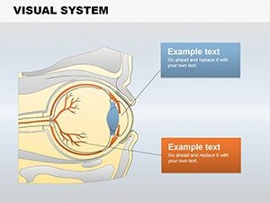
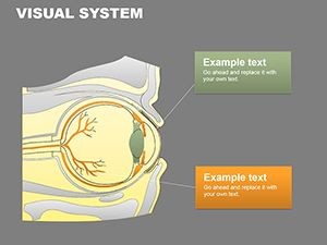
The visual system is foundational to perception, and this template, under Medicine - Pharma, captures its essence. Use shapes to diagram how the sclera protects while the cornea focuses light, ideal for pharma reps explaining drug impacts on eye health. Editable elements allow zooming into the iris's color variations or pupil adjustments, supporting discussions on conditions like glaucoma. In academia, it aligns with curricula on sensory biology, referencing studies from journals like Nature Neuroscience.
Highlighted Features for Medical Precision
- 14 Editable Slides: Comprehensive coverage from overall eye structure to detailed parts.
- Customization Options: Modify sizes, colors, and labels for tailored explanations.
- Anatomy-Focused Shapes: Accurate depictions of vessels, lenses, and holes like the pupil.
- Keynote Compatibility: Native .key format for Apple users, with export capabilities.
- Educational Aids: Built-in annotations for step-by-step breakdowns.
Superior to generic shapes, it ensures medical-grade accuracy.
Medical and Educational Use Cases
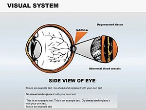
A doctor might use these shapes in patient education: A slide showing choroid blood vessels to explain nutrition's role in eye health. Pharma examples include illustrating drug delivery to the retina, as in treatments for macular degeneration per FDA approvals. Educators can create interactive lessons on refraction, engaging students with dynamic pupil simulations. Researchers reference it in papers, akin to visuals in PubMed articles. Versatile for telemedicine or conferences.
Workflow for Custom Anatomy Shapes
- Pick a Base: Select from holistic or focused eye views.
- Label Components: Add terms like 'cornea' with precision.
- Adjust for Clarity: Scale or color-code for emphasis.
- Incorporate Data: Overlay stats on light refraction.
- Present Effectively: Use transitions for revealing layers.
This approach aids in clear, impactful delivery.
Tips for Anatomical Presentations
Enhance with real scans integration, per radiology best practices. Use gradients for depth, mimicking 3D models. Cite sources like Gray's Anatomy for authority. For audiences, simplify jargon - explain pupil as 'light regulator.' Users find it boosts retention, aligning with cognitive load theory. Outperforms basic Keynote tools with specialized designs.
Illuminate vision science with confidence.
Enhance Your Visual Explanations
This template is key to precise communication. Ready to see clearly? Download and innovate.
Frequently Asked Questions
How accurate are the shapes?
Based on standard anatomy, fully editable for specifics.
Compatible with other software?
Primarily Keynote, exportable to PDF or images.
Suitable for pharma training?
Yes, ideal for drug-eye interaction visuals.
Can I add animations?
Keynote supports easy additions for dynamics.
What if I'm not a medical expert?
Intuitive for all, with built-in guides.