Tomography Medical Documentation Word Template: Clarity in Cross-Sections
Type: Word templates template
Category: Medicine - Pharma
Sources Available: .dotx, .jpg
Product ID: WT01512
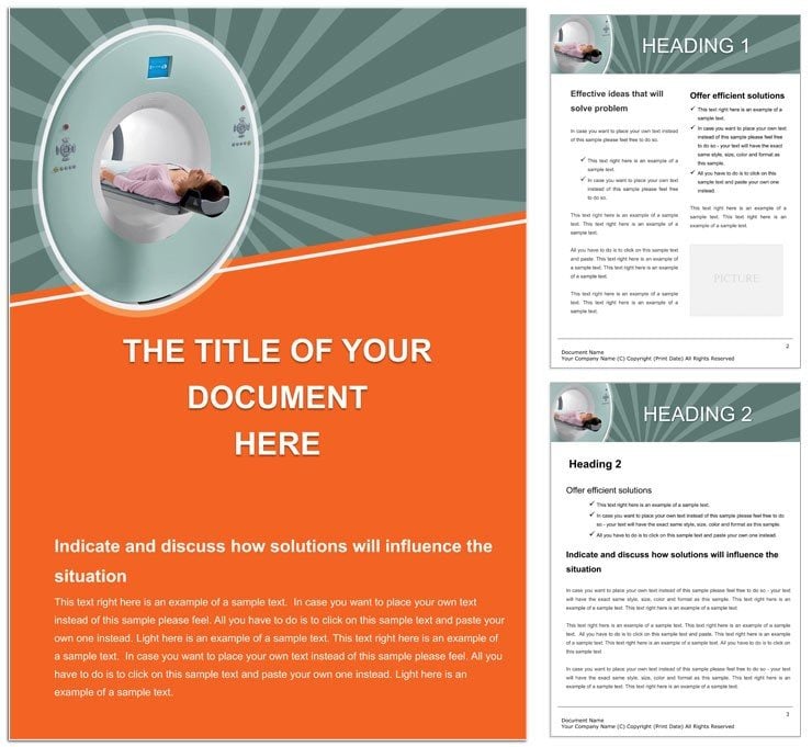
Tomography's narrow-beam precision reveals the body's hidden layers, and your documentation should match that focus. Our Tomography Medical Documentation Word Template equips radiologists, pharma researchers, and clinicians with a framework for articulating axial views of organs - from lung slices to bone densities. In the medicine-pharma realm, where details dictate decisions, this tool organizes findings into logical flows: patient history, scan parameters, image annotations, and conclusions. Built for Microsoft Word with .dotx files, tweak tables for measurements or embed placeholders for scans, ensuring reports read like diagnostic roadmaps. At $22, it's a cornerstone for accurate, efficient records that support everything from treatment plans to research papers. Whether deciphering CT contrasts or MRI gradients, this template distills complex data into accessible narratives, aiding teams in swift, informed actions.
Technical Features for Diagnostic Depth
Engineered for accuracy, it includes grid tables for coordinates, caption styles for figure refs, and bullet schemas for anomaly lists, all scalable to case complexity.
- Scan-Specific Layouts: Sections for beam angles, slice thicknesses, and contrast agents, with formulas for quick calcs.
- Image Integration: Frames for inserting axial previews, labeled with axes for orientation.
- Compliance Aids: Footers for dates/signatures, headers for case IDs, aligning with pharma standards.
- Flexible Formatting: Color-code risks - red for urgents - while keeping text neutral for prints.
Such structure sharpens insights; a researcher used it to catalog organ volumes, streamlining grant submissions.
Applications Across Imaging Fields
Radiologists draft routine reports, populating grids with Hounsfield units for tissue diffs. Pharma teams document trial scans, with expandable appendices for raw data dumps.
In oncology, outline tumor margins with annotated sketches; for orthopedics, chart fracture planes. Academic users build teaching modules, layering explanations over basics.
Precise Editing Protocol
- Initiate the Doc: Load .dotx; outline view shows hierarchy for targeted fills.
- Detail the Scan: Enter params in tables - use merge cells for multi-slice summaries.
- Annotate Visuals: Paste images; add callouts via text boxes for key findings.
- Audit Accuracy: Cross-check units; format for legibility in shared views.
- Finalize Flow: Export or save as template, versioning for ongoing series.
This keeps reports rigorous yet readable.
Strategies for Superior Scans
Use conditional formatting for auto-highlights on abnormals. Landscape for panoramic slices, portrait for narratives. Over generic forms, this's specificity accelerates reviews - a tech noted faster peer validations.
Hyperlink to protocols, embedding knowledge. For pharma, tag with trial phases for traceability.
Precision documented: layers unlocked.
Focus Your Findings
Illuminate diagnostics - download the Tomography Medical Documentation Word Template for $22 and section your success.
Frequently Asked Questions
What file type is it?
Primary .dotx for Word, with JPG previews for quick refs.
Can I insert actual scan images?
Yes, dedicated slots handle embeds, resizing to fit without crops.
Suitable for research reports?
Ideal - appendices and refs support detailed pharma studies.
How to add measurements?
Built-in tables with columns for dimensions, auto-summing where needed.
Compatible with other software?
Exports to PDF for universal viewing; core in Word.