Immune System Diagrams PowerPoint Template - Fully Editable & Instant Download


Type: PowerPoint Charts template
Category: Medicine - Pharma, Pie
Sources Available: .pptx
Product ID: PC00171
Template incl.: 30 editable slides
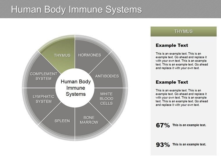
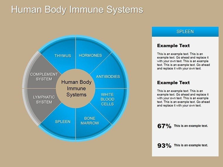
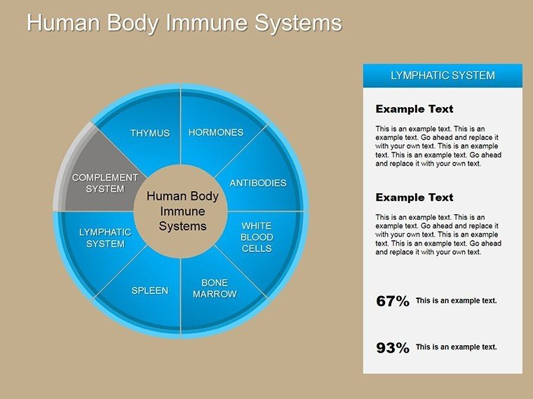
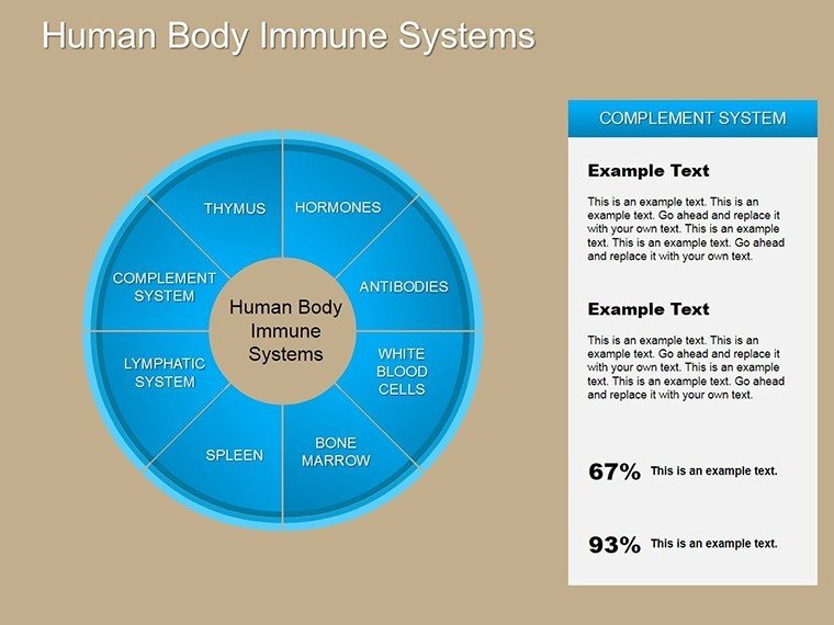
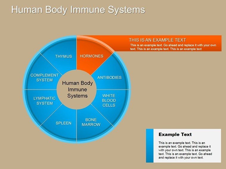
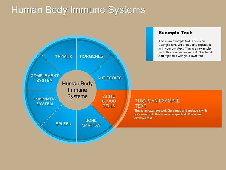
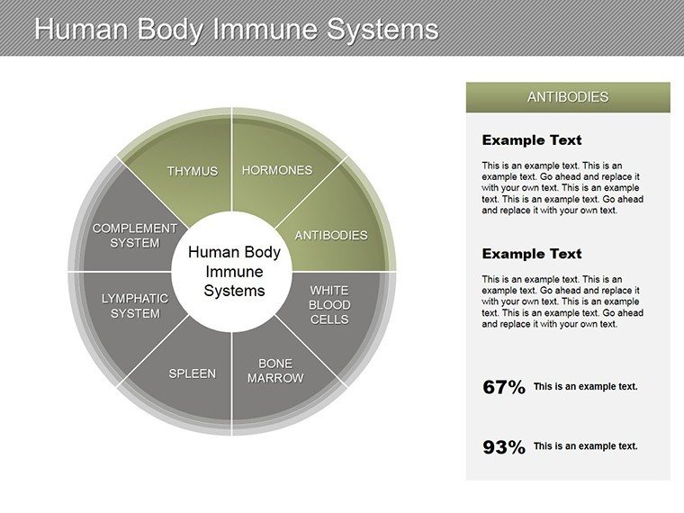
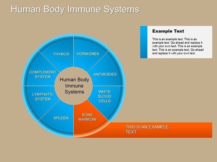
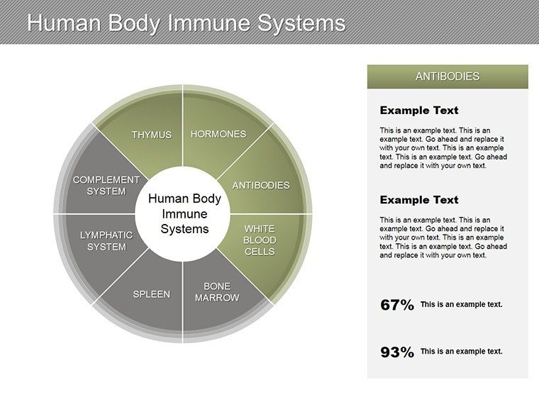
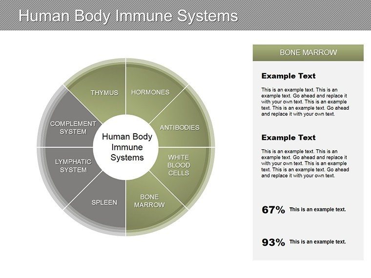
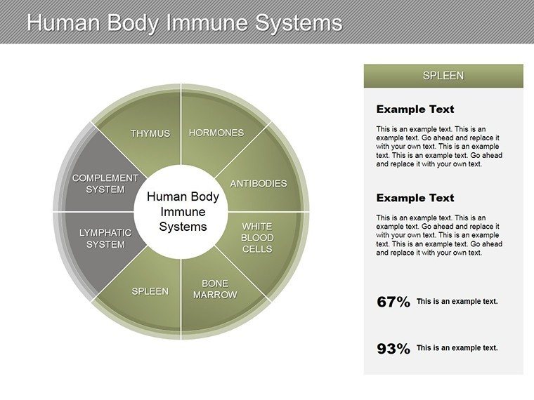
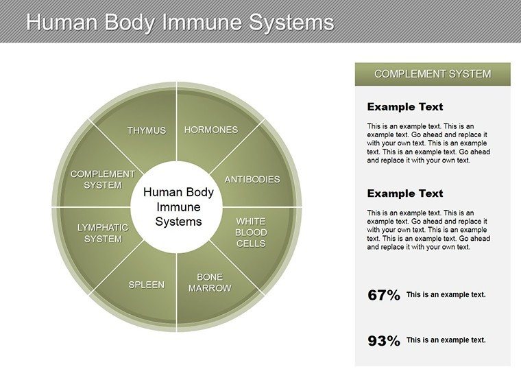
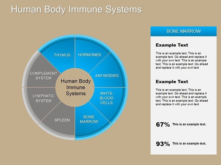
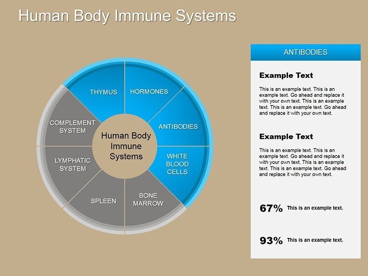
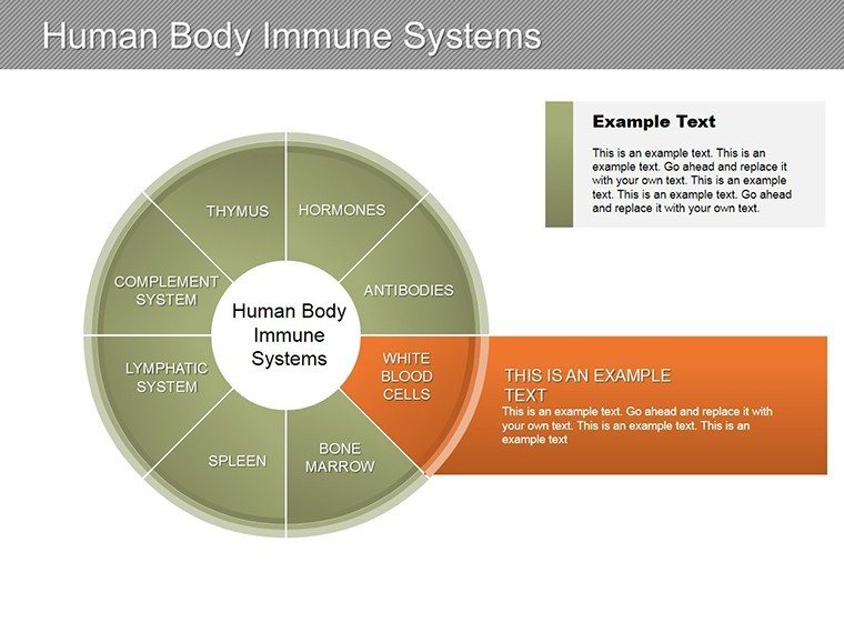
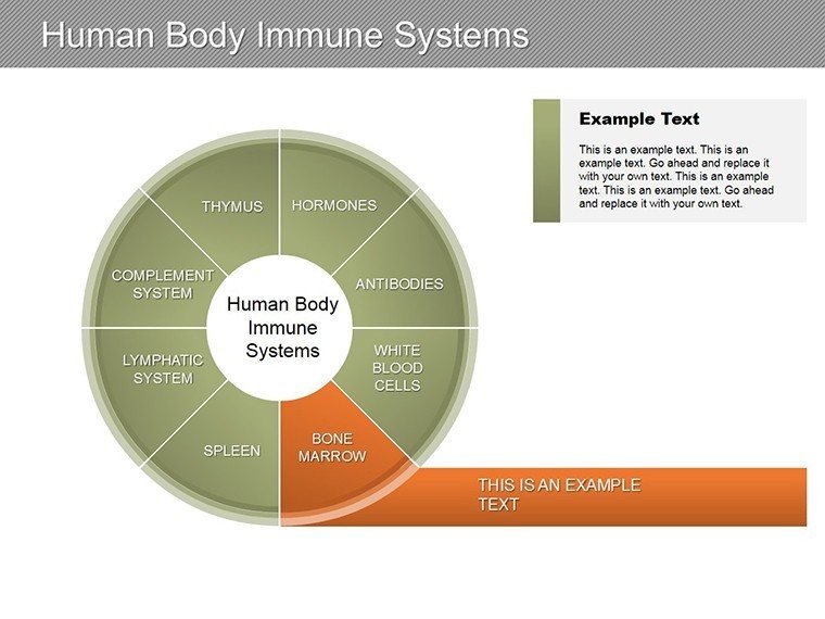
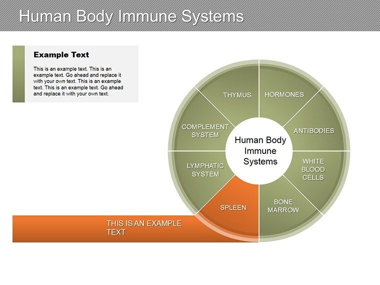
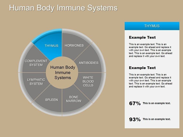
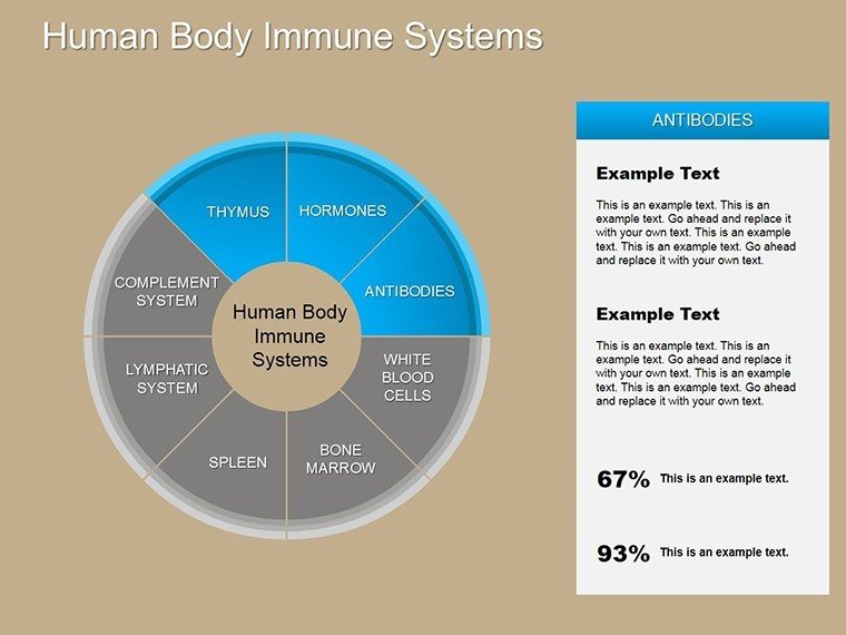
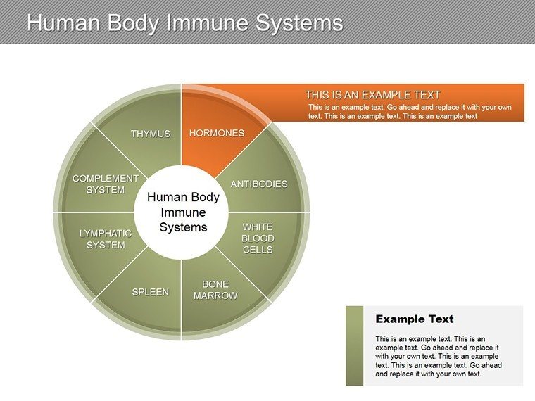
Present complex immunology concepts with clarity using this fully editable Immune System Diagrams PowerPoint Template. Designed for educators, healthcare professionals, and students, it includes professional charts that simplify explanations of the body's defense mechanisms and immune responses.
Key Features
- Fully editable vector diagrams for complete customization
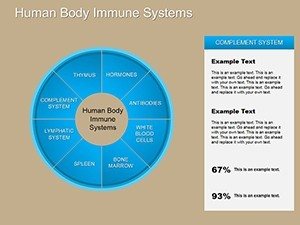
- Comprehensive coverage of innate and adaptive immunity
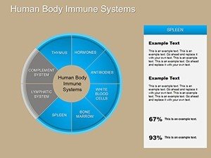
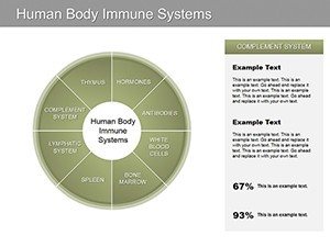
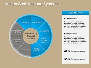
- Detailed visuals of white blood cell types and roles
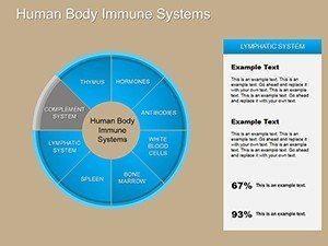
- Clear flowcharts for lymphatic system and pathways
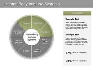
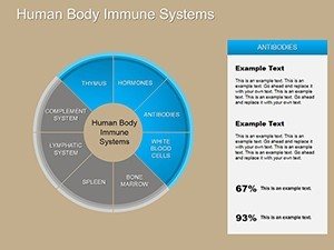
- Illustrations of antigen-antibody interactions
- Timelines depicting stages of immune responses
Benefits of This Template
This template allows you to create visually appealing presentations quickly, without needing advanced design skills.
The accurate and professional diagrams ensure your content is both informative and engaging, helping your audience grasp difficult concepts more easily.
By using ready-made charts, you can focus on your message rather than on creating graphics from scratch.
How to Use the Immune System Diagrams
Download the template and open it in Microsoft PowerPoint.
Choose the relevant diagrams and integrate them into your slides.
Easily modify text, colors, and layouts to align with your presentation style or branding.
Apply animations to demonstrate processes step-by-step for greater impact.
Professional Scenarios Where This Template Excels
This versatile template is ideal for a variety of settings:
- University lectures on biology and immunology
- Continuing education sessions for nurses and doctors
- Research symposiums and conference presentations
- Patient education in medical practices
- High school or college student projects and reports
Enhance your next presentation with these high-quality immune system diagrams - download today.
Frequently Asked Questions
Is this template fully editable in PowerPoint?
Yes, every element including shapes, text, colors, and layouts is fully editable, giving you complete control over the design.
What specific immune system topics are covered?
The template features diagrams on innate and adaptive immunity, white blood cell functions, lymphatic system flows, antigen-antibody interactions, and immune response timelines.
Is this template compatible with Google Slides?
Yes, you can upload the .pptx file to Google Slides and edit it there, though some advanced features may work best in Microsoft PowerPoint.
Do I need any special software to use this template?
No, it works with standard Microsoft PowerPoint. No additional plugins or fonts are required.
Can I use this template for commercial or educational purposes?
Yes, once downloaded, you can use it for both personal and commercial presentations without restrictions.