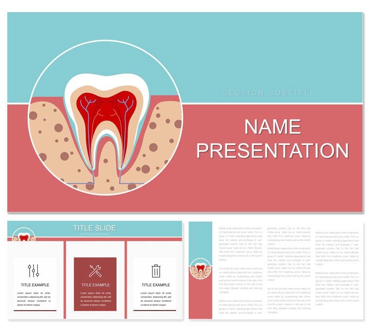
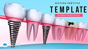
Unlock the intricate world beneath the surface of a smile with visuals that reveal layers as effortlessly as a skilled hygienist. The Tooth Anatomy Keynote template provides 59 slides dedicated to demystifying dental architecture, ideal for educators, researchers, or clinicians aiming to illuminate structures from crown to root. In Keynote, it becomes your precision instrument, blending scientific accuracy with visual elegance.
Designs peel back enamel like an onion, using stratified colors to denote dentin depths and pulp vitality, against backgrounds that evoke lab sterility or classroom curiosity. Three masters standardize anatomical labels, with three options for contextual shifts - detailed for lectures, simplified for overviews. Portable across Apple ecosystems, it facilitates annotations during study sessions or live dissections.
Precision Features for Anatomical Depth
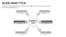
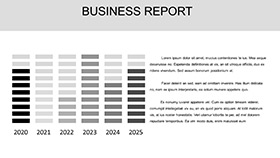
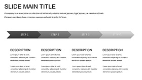
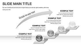
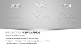
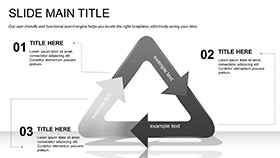
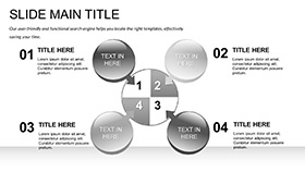
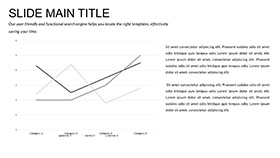
Twenty-eight diagrams anchor the template, such as exploded views segmenting incisor components, with connectors tracing nerve pathways for interactive exploration. Infographics map eruption timelines, bubbles denoting milestones like first molars emerging.
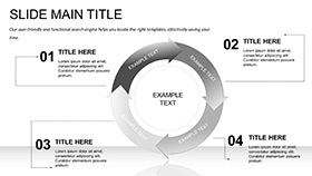
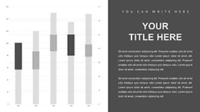
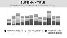
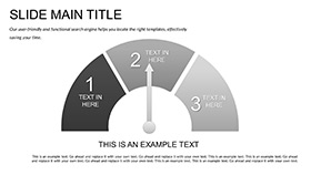
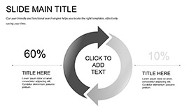
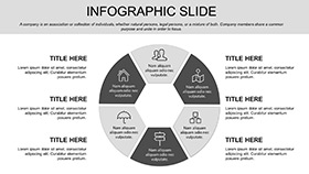
Seven schemes modulate from stark anatomical whites to illustrative pastels, suiting diagrams that evolve with user inputs. Tables catalog tissue types, columns for properties like hardness scales, while charts radialize functions around central cusps.
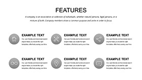
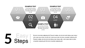
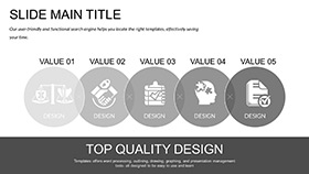
Elements Built for Discovery
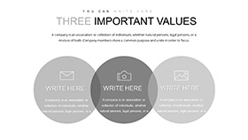
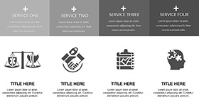
- Layered Icons: Peelable vectors of enamel and cementum, revealing subsurface on click.
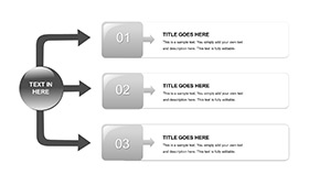
- Modular Sections: Grids for cross-sectional inserts, aligning multiple views cohesively.
- Annotation Frames: For glossaries or sourced diagrams, enhancing with hyperlinks to references.
These tools expedite complex renders, focusing energy on interpretive insights.
Anatomy in Action Across Fields
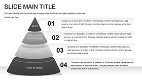
For med students, sequence diagrams guide virtual dissections, arrows directing from gingiva to apex, fostering retention through guided paths. In forensic contexts, comparative overlays highlight wear patterns, aiding case reconstructions.
Layering Your Custom Anatomy
- Frame the Focus: Choose a master suiting your scale, from macro jaws to micro tubules.
- Assemble Layers: Populate with labels; tint schemes to differentiate live vs. decayed states.
- Enrich with Details: Embed micrographs in frames, scaling for magnification effects.
- Iterate Interactivity: Add Keynote zooms to burrow into pulp chambers.
- Present Layer by Layer: Animate builds to simulate erosion, engaging learners kinesthetically.
Such steps yield decks that dissect without dismay.
Advancing Past Basic Blueprints
Surpassing sketchy sketches, it offers rotatable 3D proxies and metric-aligned scales for authenticity. SVG exports preserve fidelity for journals, with alt-text readiness for accessibility.
A bio prof leveraged it for a grant proposal, flowcharting evolutionary tooth adaptations; the clarity swayed reviewers. Versatile for textbooks or telehealth, it elevates every exposition of enamel enigmas.
Dissect Excellence in Your Decks
Core into clarity - acquire the Tooth Anatomy Keynote template at $22 and structure sessions that resonate to the root.
Frequently Asked Questions
How detailed are the structure diagrams?
They cover layers from enamel to periodontal ligament, with scalable details for various expertise levels.
Can diagrams be rotated in Keynote?
Yes, vector elements support full rotation and scaling without quality loss.
Is it geared for beginners?
It includes simplified overviews alongside advanced views, toggleable for audience adaptation.
What about nerve pathway visuals?
Dedicated flow lines trace innervations, customizable for emphasis on pain points.
Supports 3D elements?
Pseudo-3D layers simulate depth, integrable with Keynote's perspective tools.