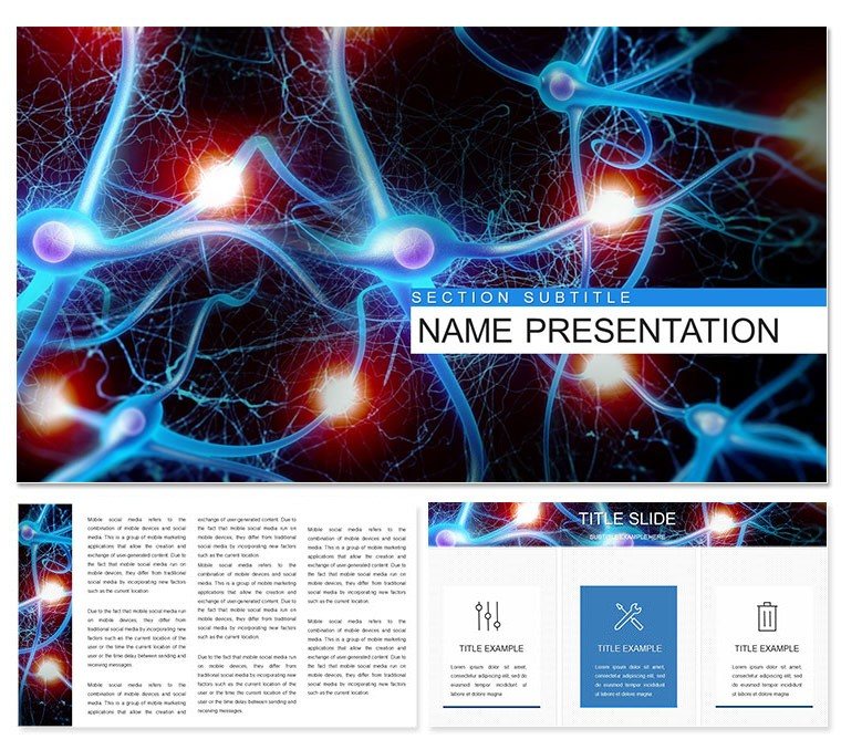
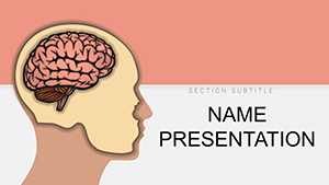
The human nervous system is a marvel of intricate pathways, much like the slides in this Keynote template that make unraveling it feel less like dissection and more like discovery. Tailored for medical students, educators, and clinicians aiming to illuminate the brain's command center, this collection of 28 diagrams transforms abstract anatomy into accessible insights. From neuron firings to spinal relays, each visual invites you to trace signals as if holding a scalpel and a spotlight.
What sets this apart? It's the thoughtful blend of detail and digestibility. Three master slides anchor your lecture, with backgrounds evoking neural webs in soft grays and synaptic blues. Seven color schemes adapt to themes - vibrant for student hooks, muted for journal clubs. Fully editable in Keynote, layer in your annotations or swap sketches for scans, all while preserving the flow. Whether prepping for exams or patient consults, this template equips you to convey how nerves orchestrate everything from reflexes to recollections.
Core Elements for Neural Narratives
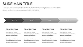
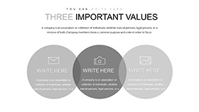
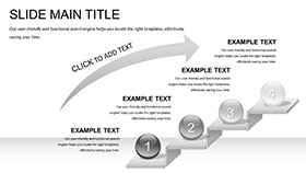
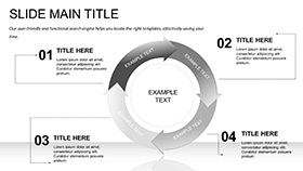
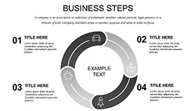
Foundationally, the three masters support varied formats: overview hubs, deep-dive panels, and recap grids. The 28 diagrams dissect systematically - cross-sections of the brainstem, 3D-ish renders of lobes, and flow models for autonomic responses. Icons of axons and dendrites are precise, ready for magnification without pixelation, and paired with callouts for functions like "motor output."
- Interactive Builds: Animate action potentials zipping along fibers, mimicking real transmission.
- Note Integration: Embed explanatory text boxes that expand on slide, like pop-up glossaries.
- Palette Precision: Seven options to differentiate central from peripheral, or highlight pathologies in cautionary reds.
These aren't mere illustrations; they're scaffolds for understanding. A professor might use a layered diagram to peel back meninges, revealing ventricles step by step.
Dissecting the Diagram Suite
Organize by region: Central nervous slides feature brain atlases with labeled gyri, peripheral ones map dermatomes via sensory grids. Physiology gets dynamic - waveforms for EEG patterns or loops for feedback in homeostasis. All primed for Keynote's data tools: Link to spreadsheets for reflex arc timings.
- Annotate freely: Draw arrows on neuron types, labeling multipolar versus bipolar.
- Sequence reveals: Build synapse gaps gradually, syncing to your discourse on neurotransmitters.
- Adapt for handouts: Flatten animations to static views for study guides.
This approach aligns with curricula from sources like Gray's Anatomy, prioritizing layered revelation.
Applications in the Classroom and Clinic
Consider a neuroanatomy seminar: Launch with this template, embedding fMRI slices on a cortex map, animated to light up during tasks. Students trace pathways, grasping Broca's role without rote memorization.
For grand rounds, employ cycle diagrams for pain signaling, customizing hues to flag chronic versus acute. A resident adjusts nodes for case specifics, fostering dialogue on interventions. It's elucidation at work, where visuals bridge theory to triage.
Extend to patient education: Simplified versions outline migraine triggers via branching trees, empowering discussions on management. Or in workshops, radial charts fan out disorders - from Alzheimer's plaques to MS demyelination - equipping attendees to navigate nuances.
Blending with Study Tools
Sync with Anki for flashcard exports or Visible Body apps for 3D spins - diagrams serve as static anchors. Insider tip: Use slide transitions to simulate nerve impulses, adding rhythm to recaps.
Open strong: A macro of a firing neuron, pulsing to hook curiosity from the get-go.
Superior to Sketchpad Basics
Outshines generic slides with medically attuned proportions - no distorted lobes here. It's like a digital Netter's - concise yet comprehensive. Educators value the note fields, turning decks into self-contained modules for flipped classrooms.
Go further: Hyperlink regions to PubMed abstracts, or script builds for quiz interactions. Typography favors sans-serifs for legibility under lecture hall lights.
Tailoring Tricks
- Scheme sync: Match colors to histological stains for authenticity.
- Layer logic: Isolate elements for focus, like solo spinal cords.
- Export ease: Generate variants for online modules versus in-person.
Wire up your next lesson - secure this template and spark synaptic connections.
Frequently Asked Questions
Compatible Keynote versions?
Works smoothly from 2016 onward, across macOS updates.
Diagram total?
28, from gross anatomy to functional models.
Custom notes possible?
Yes, dedicated fields for detailed expansions.
Animation for physiology?
Built for it - customize to depict signal propagation.
Format options?
.key for full use, .kth for theme imports.
Group study fit?
Perfect - cloud-enabled for collaborative annotations.